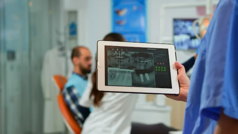
Dental radiography stands as a pivotal tool in modern dentistry, particularly in the areas of diagnosis and treatment planning. This imaging technology enables dentists to view the hidden areas of the mouth that are not visible during a regular dental examination. By using dental X-rays, various conditions such as cavities, gum disease, and infections can be detected early, enhancing the effectiveness of treatments. Moreover, dental radiography is crucial in planning complex procedures like implants and orthodontic treatments, where precision is paramount to successful outcomes. Thus, understanding the capabilities and applications of dental radiography can significantly impact patient care in dentistry.
Importance of Dental Radiography
Dental radiography, commonly known as dental X-rays, plays a crucial role in the field of dentistry. This diagnostic tool allows for the detailed visualization of the teeth, bones, and surrounding tissues that are not visible during a standard dental examination. By leveraging the technology of radiography, dentists can provide more accurate diagnoses and effective treatment plans. The importance of dental radiography cannot be overstated. It aids in the early detection of dental issues, including tooth decay, bone loss, and abscesses, which can be identified before becoming severe. Additionally, dental radiographs offer an assessment of dental health, helping in the evaluation of the condition of existing dental work and the planning of future procedures like implants and orthodontic treatments.
Early Detection of Dental Issues
One of the primary benefits of dental radiography is its capability for early detection. Dental X-rays can reveal hidden dental structures, such as wisdom teeth, and detect early signs of dental caries (cavities) that are not visible during a typical dental examination. Identifying cavities at an early stage allows for less invasive treatments and can prevent the progression of decay that would require more complex procedures.

Furthermore, dental radiography can help in the detection of bone loss associated with periodontal disease. Periodontitis often progresses without obvious symptoms until significant damage has occurred. By using radiographs, dentists can measure bone levels and detect bone loss early, enabling timely intervention to halt or reverse the condition.
Another critical aspect is the identification of dental abscesses and cysts. These conditions can be extremely painful and may lead to more serious health issues if not treated promptly. Radiographs provide a clear image of these infections, often before symptoms become severe, allowing for early intervention and improved patient outcomes.
Assessment of Dental Health
Dental radiography is indispensable for a comprehensive assessment of dental health. It helps in evaluating the condition of existing dental work, such as fillings, crowns, and bridges. Assessing the integrity of these restorations ensures they are functioning correctly and not contributing to further dental issues.
In the context of implantology and bone regeneration, radiographs are particularly vital. They help in assessing the quality and quantity of bone available for implant placement. This evaluation is crucial for successful dental implant procedures and ensures that the implant will have sufficient support for long-term stability.
Dental radiographs also aid orthodontists in planning orthodontic treatments. By examining the position and growth of teeth, radiographs help in devising effective treatment plans for moving teeth into their desired positions. This improves not only the function but also the aesthetics of the patient’s smile.
Overall, dental radiography is an essential tool in modern dentistry. Its ability to reveal hidden issues and provide a thorough assessment of dental health helps in delivering high-quality care to patients. For further insights into dental technologies and treatments, feel free to explore our other articles.
Types of Dental Radiographs
Dental radiographs, commonly referred to as X-rays, are an essential diagnostic tool in modern dentistry. They allow for the visualization of structures within the mouth and jaw that are not visible to the naked eye. Radiographs help in diagnosing, monitoring, and planning treatment for a variety of dental conditions. Understanding the different types of dental radiographs is crucial for both patients and practitioners to ensure optimal dental care.
There are several types of dental radiographs, each serving a distinct purpose. The two primary categories are intraoral and extraoral radiographs, with specific subtypes within each category. Each type provides unique information that is critical for accurate diagnosis and treatment planning. Below, we will explore these categories in more detail, along with specific examples such as bitewing radiographs.
Intraoral Radiographs
Intraoral radiographs are taken with the X-ray film placed inside the mouth. These are the most common type of dental X-rays, providing a high level of detail. Intraoral radiographs allow dentists to detect cavities, examine the tooth roots, check the health of the bony area around the tooth, and monitor the status of developing teeth.
There are several types of intraoral radiographs, including:
- Periapical X-rays – focus on showing the entire tooth, from the crown to the root.
- Bitewing X-rays – typically used to detect decay between the teeth and changes in bone density caused by gum disease.
- Occlusal X-rays – capture the floor of the mouth to show the development and placement of an entire arch of teeth.
Intraoral radiographs are invaluable for preventive care and early intervention. By identifying issues early, dentists can provide treatments that prevent further damage and maintain oral health.
Extraoral Radiographs
Extraoral radiographs are taken with the X-ray film outside the mouth. These images provide information about the larger structures of the jaw and skull. While they do not offer the same level of detail as intraoral X-rays, they are crucial for evaluating the overall condition of the jaw and facial bones.
Examples of extraoral radiographs include:
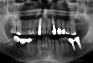
- Panoramic X-rays – show a broad view of the jaws, teeth, sinuses, nasal area, and temporomandibular joints (TMJ).
- Cephalometric projections – used primarily in orthodontics to assess the relationships between teeth and jawbones.
Extraoral radiographs are particularly useful in the planning and monitoring of orthodontic treatments, evaluating TMJ disorders, and assessing the need for procedures like dental implants and sinus lifts.
Bitewing Radiographs
Bitewing radiographs are a type of intraoral X-ray specifically designed to capture the crowns of the upper and lower teeth. They are called “bitewing” because the patient bites down on a wing-shaped device that holds the X-ray film in place. This type of radiograph is crucial for identifying decay between teeth and changes in bone density that may indicate periodontal disease.
Bitewings are typically taken during routine dental check-ups and are essential for detecting early signs of dental decay. They allow dentists to identify cavities that are not visible during a clinical examination, particularly those that occur between the teeth.
The benefits of bitewing radiographs include:
- Early detection of cavities in areas not visible during a clinical exam.
- Monitoring changes in bone density that can indicate periodontal disease.
- Assessing the fit of dental restorations like crowns and bridges.
Understanding the importance of bitewing radiographs can help patients appreciate the value of regular dental check-ups. By catching issues early, these radiographs contribute to long-term oral health and prevent more extensive dental problems.
For more detailed information on dental radiographs and their applications, be sure to explore our other articles. Each type of radiograph has unique benefits and uses that contribute to maintaining optimal oral health.
Procedure and Safety Considerations
When it comes to dental implantology and bone regeneration, the procedure and safety considerations are paramount. Dental professionals must adhere to stringent protocols to ensure that both the procedure and the materials used are safe for the patient. This includes the use of dental radiographs, which are essential for accurate diagnosis and treatment planning. In this section, we will explore how dental radiographs are taken, the safety measures implemented for patients, and the associated risks and benefits of radiation exposure in dental settings. Understanding these aspects can help both practitioners and patients feel more confident about the process.
How Dental Radiographs are Taken
Dental radiographs, commonly known as X-rays, are a crucial diagnostic tool in implantology and bone regeneration. The process involves placing a small sensor inside the patient’s mouth, which captures images of the teeth, bone, and surrounding tissues. These images provide valuable information that helps in planning the treatment accurately.
There are several types of dental radiographs, including bitewing, periapical, and panoramic X-rays. Each type serves a specific purpose:
- Bitewing X-rays: Show the upper and lower teeth in a single view, often used to detect cavities between teeth.
- Periapical X-rays: Provide a detailed image of one or two teeth from crown to root, useful for detecting abnormalities in the tooth or surrounding bone.
- Panoramic X-rays: Offer a broad view of the jaws, teeth, and nasal area, essential for comprehensive treatment planning.
The procedure is quick, usually taking only a few minutes. Patients are asked to remain still while the X-ray is taken to ensure a clear and accurate image.
Safety Measures for Patients
Ensuring the safety of patients during dental radiographic procedures is a top priority. Protective measures are in place to minimize exposure to radiation and increase patient comfort. One of the most common safety measures is the use of a lead apron, which shields the body from unnecessary radiation.
In addition to the lead apron, patients may also wear a thyroid collar to protect the thyroid gland, which is particularly sensitive to radiation. Modern dental X-ray machines are designed to focus the radiation precisely, further reducing exposure risk.
Dental professionals are trained to take the minimum number of X-rays required to diagnose and plan treatment effectively. By adhering to the ALARA principle (As Low As Reasonably Achievable), they ensure that radiation exposure is kept to a minimum while still obtaining the necessary diagnostic information.
Radiation Exposure and Risks
While dental radiographs are an invaluable tool, it is essential to understand the potential risks associated with radiation exposure. Although the level of radiation used in dental X-rays is relatively low, it is still critical to consider the cumulative effect of multiple exposures over time.
Studies have shown that the risks of dental X-rays are minimal when safety protocols are strictly followed. The benefits of accurate diagnosis and effective treatment planning usually outweigh the potential risks. However, certain populations, such as pregnant women and young children, may require special considerations to further minimize exposure.
Patients with a history of extensive X-ray exposure should inform their dentist so that alternative diagnostic methods can be considered if necessary. Understanding the balance between the risks and benefits of dental radiographs is key to making informed decisions about oral healthcare.
By staying informed about the procedures and safety measures involved in dental radiography, patients can approach their treatment with greater confidence. For more detailed information on related topics, consider reading our other articles on advanced dental procedures and patient safety.
Interpreting Dental Radiographs
Dental radiographs, commonly known as X-rays, are an essential diagnostic tool in modern dentistry. They provide a detailed view of the teeth, bones, and surrounding tissues, which are not visible during a regular dental examination. Understanding how to interpret these images accurately is crucial for diagnosing and treating various dental conditions effectively.
The interpretation of dental radiographs involves analyzing the images to detect abnormalities, dental caries, periodontal diseases, and other conditions. Accurate interpretation requires a thorough understanding of both normal anatomical structures and pathological changes. Utilizing radiographs can help in planning treatments, monitoring disease progression, and ensuring the success of dental interventions.
Interpreting dental radiographs is a skill that evolves with practice and experience. Dentists and dental specialists must stay updated with the latest advancements in radiographic technology and interpretation techniques to provide the best care for their patients.
Common Findings on Dental Radiographs
Dental radiographs can reveal a variety of common findings that are crucial for diagnosing dental issues. One of the most frequent discoveries is dental caries, commonly known as cavities. These appear as darkened areas on the radiograph where the enamel and dentin have been demineralized by bacterial activity.
Another common finding is periodontal disease, which affects the supporting structures of the teeth. This can be seen on radiographs as bone loss around the teeth, indicating the severity and extent of the disease. Furthermore, impacted teeth, which are teeth that have failed to emerge fully into the dental arch, can be easily identified on radiographs.
Radiographs also help in identifying root canal infections. These appear as radiolucent areas around the root apex, indicating the presence of infection or abscess. Additionally, pathological lesions such as cysts, tumors, and other bone abnormalities can be detected and investigated further.
In summary, the common findings on dental radiographs include:
- Dental caries
- Periodontal disease
- Impacted teeth
- Root canal infections
- Pathological lesions
Role of Dentists in Interpretation
The role of dentists in interpreting dental radiographs is critical for accurate diagnosis and treatment planning. Dentists are trained to identify and differentiate between normal anatomical structures and pathological changes. This expertise is particularly important in early detection and prevention of dental diseases.
Dentists are also responsible for explaining radiographic findings to patients. Clear communication helps patients understand their dental conditions and the rationale behind proposed treatments. This transparency fosters trust and encourages patients to participate actively in their dental care.
Moreover, dentists must ensure that radiographs are taken using the lowest possible radiation dose, following the ALARA (As Low As Reasonably Achievable) principle. Dentists should continually update their knowledge and skills in radiographic techniques and interpretation through continuing education and training.
In conclusion, the role of dentists in interpreting dental radiographs involves:
- Accurate diagnosis of dental conditions
- Clear communication with patients
- Ensuring patient safety
- Continual professional development
Interpreting dental radiographs effectively allows dentists to offer comprehensive care and enhances the overall health and well-being of their patients. For further information on related topics, explore our other articles on dental health and advanced treatments.
Understanding Dental Radiography
Dental radiography plays a crucial role in diagnosing and planning treatment in the field of dentistry. Below is a common question that sheds light on its significance.
How does dental radiography assist in dental diagnosis and treatment?
Dental radiography, or dental X-rays, are essential tools for dental professionals. They provide valuable information that is not visible during a regular dental exam. These images help dentists detect hidden dental abnormalities, such as cavities between teeth, jaw bone loss, and impacted teeth. They also aid in planning treatments for dental implants, checking the results of an infection, and evaluating the status of developing teeth. Essentially, dental radiographs make it possible to diagnose and treat conditions that could otherwise go unnoticed and become more problematic.

My name is Salman Kapa, a 73-year-old expert in bone regeneration and dental implantology. With decades of experience in the field, I am dedicated to advancing our understanding of oral health and hygiene. Through my research and writing, I aim to contribute to the development of innovative solutions in dental care.


